
Morton’s Neuroma: Causes, Diagnosis, Treatment & Surgery
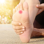
Morton’s neuroma is a painful thickening around one of the small nerves that run between the long bones of the forefoot, most often in the space leading to the third and fourth toes. It typically causes burning pain in the ball of the foot, tingling or numbness in two neighbouring toes, and the odd sensation of a pebble or folded sock under the foot.
Despite its name, it is not a tumour. It is a nerve that has been squeezed and irritated, step after step, until it has scarred and swollen. That is good news: most people improve without an operation, and when surgery is needed, the results are well studied.
This guide explains how the condition develops, how clinicians confirm it, every main treatment option from injections to newer ablation techniques, and, in detail, what surgery involves, what results to expect and what can go wrong. If you are mainly looking for footwear support, see our separate page on insoles for Morton’s neuroma.
What is Morton's neuroma?
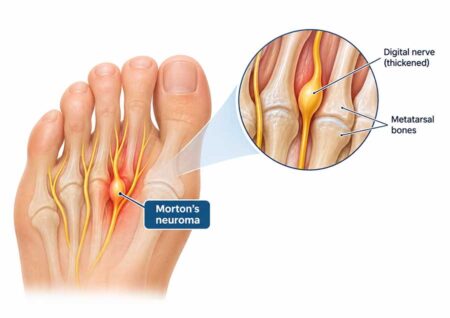
Morton’s neuroma: compression of a common digital plantar nerve (the nerve fibers divide to supply adjacent opposing toes). Repeated trauma to the nerve causes the surrounding tissue to thicken and become fibrotic, forming a firm, spindle-shaped swelling just before the nerve divides.
The swelling most often occurs in the “third web space” between the heads of the third and fourth metatarsal and less often between the second and third. Having this in two web spaces of the same foot (bilaterally) at the same time is rare and causes physicians to be more suspicious of another cause in those situations.
It is referred to by different names in the medical literature: interdigital neuroma, intermetatarsal neuroma, Morton’s metatarsalgia, and plantar digital neuritis. Neuroma is an old term. A true neuroma is a growth of nerve cells; this is fibrous scar tissue around a chronically irritated, normal-sized nerve.
A short history
The condition was first described by a London chiropodist, Lewis Durlacher, in 1845, and later by the American surgeon Thomas George Morton in 1876, whose name stuck. In 1951 the British surgeon J.D. Mulder described the “click” that is still used in clinic today.
Who gets Morton’s neuroma?
It mainly affects adults in middle age, and women far more than men. In a UK study of 253 family practices covering 1.83 million patient-years, new diagnoses of Morton’s metatarsalgia were made at a rate of 87.5 per 100,000 women a year, compared with 50.2 per 100,000 men, with the peak between ages 55 and 64 (Latinovic et al., 2006).
The same study shows how rarely it needs an operation: only about 3% of newly diagnosed patients had surgery that year. Most people manage it with simpler measures.
It is also common to have a neuroma without knowing it. MRI studies of people with no foot pain have found neuroma-like swellings in up to a third of them (Bencardino et al., 2000). This is why scans are matched with symptoms and examination, not read in isolation.
What does Morton’s neuroma feel like?
Most people describe a mix of nerve-type and pressure-type symptoms in the ball of the foot:
- burning, sharp or electric pain between two metatarsal heads, often shooting into the toes
- tingling, pins and needles or numbness in the two toes on either side of the affected space
- the feeling of walking on a marble, pebble or rucked-up sock
- pain that builds in tight or high-heeled shoes and eases when the shoe comes off and the forefoot is rubbed
- a click or clunk when the forefoot is squeezed
Early on, symptoms come and go and may only appear in certain shoes. Over months they can become more frequent and last longer. Pain at rest or at night is less typical and should prompt a check for other causes.
What causes Morton’s neuroma?
No single cause has been proven, but most experts see it as a repetitive-injury problem. Four explanations are discussed in the medical literature, and they probably work together.
- Entrapment. The nerve passes beneath the deep transverse metatarsal ligament, a tough band tying the metatarsal heads together. When the toes bend upward at push-off, the nerve is pulled against the front edge of that band. Repeated thousands of times a day, this rubbing is thought to scar the nerve’s outer sheath.
- Repetitive trauma. Pressure from below, especially on hard surfaces or in thin-soled shoes, compresses the nerve between the ground and the ligament.
- Reduced blood supply. Long-standing pressure may limit blood flow to the nerve, contributing to the fibrous changes found when neuromas are examined under the microscope.
- Bursitis. A small fluid sac (bursa) sits between the metatarsal heads. When it becomes inflamed and enlarged, it can push down on the nerve.
Why the third web space?
The nerve to the third space is often fed by branches of two different nerves, which can make it slightly thicker and less mobile. The third and fourth metatarsals also move differently: the inner three are relatively fixed, while the outer two are more flexible. That difference in motion is thought to add shearing stress exactly where the nerve runs.
Risk factors
- Footwear. Narrow toe boxes squeeze the metatarsal heads together; high heels shift weight forward and keep the toes bent upward, stretching the nerve against the ligament.
- Forefoot-heavy activities. Running, court sports, dance and any work that involves crouching or kneeling on the toes. In Canada, snug ski boots, hockey skates and clipped-in cycling shoes are frequent triggers because they compress the forefoot for long periods.
- Foot shape. Bunions, hammer toes, a flexible flat foot or a high, rigid arch can all change how load is shared across the forefoot.
- Tight calf muscles. Limited ankle movement makes the heel lift early, so the forefoot takes more load for longer. Calf tightness has been linked to forefoot nerve entrapment (Barrett & Jarvis, 2005).
- Sex and age. Women are diagnosed far more often, which most researchers link to footwear rather than biology alone. Risk rises through middle age as the forefoot’s fat pad thins.
How is Morton’s neuroma diagnosed?
It is mainly a clinical diagnosis: a careful history and a hands-on examination are usually enough, with a scan used when the picture is unclear or before any procedure.
The examination
Your clinician will ask where the pain is, what it feels like, which shoes or activities set it off and whether your toes go numb. They will then press between each pair of metatarsal heads and compare the two feet. The most useful tests are:
- Web space squeeze (thumb–index finger test). The examiner pinches the painful web space between thumb (underneath) and index finger (on top). Reproducing your pain is a strong sign. Compared with ultrasound, this simple test picked up 96% of neuromas, making it the most accurate of seven tests studied (Mahadevan et al., 2015). A 2024 systematic review confirmed it as highly sensitive (Pitcher et al., 2024).
- Mulder’s click. The examiner squeezes the forefoot from side to side while pressing the web space from below. A palpable click, sometimes with a jolt of pain, suggests the swollen tissue is popping between the bones. It is well known but less reliable, catching about 61% of neuromas in the same study.
- Sensation testing. Light touch or a pin on the facing sides of the two toes may show reduced feeling.
Imaging
- X-ray cannot show nerves, but it is often ordered to rule out a stress fracture, arthritis or a deformity of the joints.
- Ultrasound shows the neuroma as a dark, oval swelling between the metatarsal heads and can be done while the foot is squeezed. A meta-analysis of 14 surgically confirmed studies found ultrasound picked up 91% of neuromas and MRI 90%, with no real difference between them (Bignotti et al., 2015). Ultrasound is quicker, cheaper and widely available in Canada through hospital and community imaging clinics.
- MRI is useful when the diagnosis is uncertain, when more than one problem may be present, or before surgery. Because small neuromas show up in many people without symptoms, the size and location on the scan must match where it hurts.
Diagnostic injection
A small injection of local anaesthetic into the web space can help confirm the source. If the pain switches off for a few hours, the nerve in that space is very likely responsible. This is particularly helpful when two spaces are tender.
Conditions that can be mistaken for Morton’s neuroma
Forefoot pain has several causes, and some sit right next to the neuroma. Getting this right matters, because surgery on the wrong structure will not help.
| Condition | What sets it apart |
|---|---|
| Metatarsalgia (mechanical) | Aching directly under a metatarsal head, usually without numbness; tender on the bone, not between bones |
| Stress fracture | Swelling on top of the foot and pinpoint bone tenderness after a jump in activity; may need a repeat X-ray or MRI |
| Plantar plate tear / second toe instability | Pain under the second toe joint, often with the toe drifting or lifting; can coexist with a neuroma |
| Intermetatarsal bursitis | Fluid-filled swelling in the same space; can mimic or accompany a neuroma on ultrasound |
| Joint inflammation (e.g. rheumatoid arthritis) | Several joints swollen and stiff, often both feet, morning stiffness |
| Nerve problems further up (tarsal tunnel, lower back) | Numbness over a wider area of the sole, symptoms that do not match one web space |
These overlaps are common. In one surgeon’s series of 121 patients with a neuroma, 20% also had instability of the second toe joint that needed treating at the same time (Coughlin et al., 2002).
How is Morton’s neuroma treated without surgery?
Treatment usually follows a ladder: start with the simplest changes, and step up only if they are not enough. A 2023 Cochrane review found that the evidence for nearly every treatment, surgical or not, is still limited, so the order below reflects risk and cost as much as proof (Cochrane, 2023).
Step 1: Take the pressure off
The first step is removing what squeezes the nerve: wider, lower-heeled shoes with a roomy toe box, fewer hours in tight sports footwear, and a break from the activity that flares it. Supportive insoles with a metatarsal raise are part of this step; we cover them separately on our insoles for Morton’s neuroma page. Anti-inflammatory tablets such as ibuprofen can ease flare-ups in the short term, and calf stretching helps if your ankles are stiff.
Step 2: Corticosteroid injection
A corticosteroid (“cortisone”) injection, usually mixed with local anaesthetic, is the most common next step. Results are mixed:
- In a patient-blinded trial, people given a steroid plus anaesthetic rated their foot health significantly better at three months than those given anaesthetic alone (Thomson et al., 2013).
- A smaller double-blind trial of 41 patients found no difference between the two at three and six months, and about half of each group went on to request surgery (Lizano-Díez et al., 2017).
- Followed for almost five years, a single injection was still working for 36% of patients (Hau et al., 2021).
Injections are quick and can also confirm the diagnosis. Repeated doses carry risks: thinning of the protective fat pad under the ball of the foot and lightening of the skin, each seen in a few percent of patients in the 2013 trial. Most clinicians limit the number of injections for that reason.
Step 3: Procedures that target the nerve
If injections fail, some clinics offer treatments that damage or calm the nerve without open surgery. These are newer, mostly studied in case series, and availability in Canada varies, often through private clinics.
| Treatment | How it works | What the research shows | Watch-outs |
|---|---|---|---|
| Alcohol (sclerosing) injections | A series of dilute alcohol injections, usually under ultrasound, chemically damages the nerve | Short-term results are encouraging, but at about 5 years only 29% of 45 patients were symptom-free and 36% had gone on to surgery (Gurdezi et al., 2013). A newer stronger single-injection technique reported 82% success at 5 years in 33 feet (Pabinger et al., 2020) | Several visits; post-injection pain and flare-ups; results vary with technique |
| Radiofrequency ablation | A needle probe heats the nerve to stop it sending pain signals | A 2025 meta-analysis of 8 studies (237 patients) found large pain reductions and complications in about 2% (Llombart-Blanco et al., 2025) | Short follow-up in most studies; long-term durability unknown |
| Cryoablation | A probe freezes the nerve under ultrasound or MRI guidance | In one case series of 24 treatments, 83% of patients were very satisfied with no complications (case series, 2023) | Small numbers; nerve may regrow over time |
| Shockwave therapy (ESWT) | Pressure waves applied through the skin over several sessions | In a randomized sham-controlled trial, pain fell significantly at 1 and 4 weeks with shockwave but not with sham (Seok et al., 2016) | Very short follow-up; neuroma size did not change |
Some insurers still class alcohol injection, radiofrequency ablation and cryoablation as investigational, so check your extended health plan before booking.
Surgery for Morton’s neuroma: what you need to know
Surgery is the most reliable way to stop persistent neuroma pain, but it is a trade: most people swap pain for a patch of permanent numbness, and a minority are left with pain of a different kind. Understanding the options and the real-world results helps you make that decision with your surgeon.
When is surgery considered?
Surgery is usually offered when all three of these are true:
- symptoms have lasted several months despite proper footwear changes and at least one injection or similar treatment
- the diagnosis is clear, ideally with a positive squeeze test, an imaging finding in the same web space and relief from a diagnostic anaesthetic injection
- the pain is limiting walking, work or sport enough to justify the risks
There is rarely a rush. In a prospective study of 99 patients, the time between diagnosis and surgery made no difference to the final result (Bucknall et al., 2017), so it is reasonable to exhaust simpler options first.
The main types of operation
1. Neurectomy (removing the nerve). This is the standard operation. The surgeon finds the thickened segment of nerve, cuts it well back from the weight-bearing part of the forefoot so the cut end can retract between the muscles, and removes it. The tissue is sent to the lab to confirm the diagnosis. Because the nerve supplies feeling to the facing sides of two toes, those areas are usually left numb.
2. Decompression (releasing the ligament). Instead of removing the nerve, the surgeon cuts the deep transverse metatarsal ligament to give the nerve room, sometimes also moving the nerve to a safer position. The aim is to relieve pain while keeping feeling in the toes. In a long-term study of 39 patients treated this way, 64% had no pain at follow-up, 20% mild pain and 16% severe pain, and the authors suggested decompression as a first operation with neurectomy kept in reserve (Koti et al., 2022). Even so, two-thirds of their patients reported some numbness.
3. Variations aimed at specific problems. Some surgeons bury the cut end of the nerve inside a muscle to lower the risk of a painful stump forming, or combine a ligament release with a small shortening cut in the neighbouring metatarsal to widen the space. In one comparative series, adding the bone cut gave better results than ligament release alone (Park et al., 2013). Minimally invasive and endoscopic ligament releases are also offered in some centres, but long-term data are limited.
Top or bottom of the foot? Dorsal vs plantar incision
A neuroma can be reached through a cut on the top of the foot (dorsal) or on the sole (plantar). Neither is clearly better overall, but the trade-offs differ. A 2024 meta-analysis of eight studies and 549 neuromas found:
| Outcome | Plantar (sole) incision | Dorsal (top) incision |
|---|---|---|
| Reduced feeling after surgery | 48.5% | 62.0% |
| Tender scar | 16.7% | 6.2% |
| Nerve confirmed as neuroma in the lab | 99.3% | 97.1% |
| Overall complications | 8.8% | 12.0% (difference not significant) |
Source: Lee et al., 2024. An earlier review noted that the plantar approach tends to mean a longer wait before full weight-bearing and more wound problems, because the scar sits on a surface you walk on (Zhang et al., 2023). Most surgeons use the dorsal approach for a first operation and may prefer the plantar route for revisions.
How successful is Morton’s neuroma surgery?
Most people are satisfied, but fewer are completely pain-free than older figures suggested:
- Long-term, one surgeon: of 71 feet followed for an average of 5.8 years after dorsal neurectomy, 65% were completely pain-free. Numbness was reported in 36 feet but was bothersome in only four (Coughlin & Pinsonneault, 2001).
- Prospective, patient-reported: in 99 patients, 79% rated the result excellent or good and 10% poor or very poor; 63% were pain-free at six months (Bucknall et al., 2016).
- Ten years or more: 76% of 98 feet had an excellent or good result. All eight poor results were caused by a painful stump neuroma, and operating on several neuromas at once did worse than a single one (Kasparek & Schneider, 2013).
In the Bucknall study, obesity, depression, social deprivation and revision surgery were all linked to poorer outcomes, which is worth discussing honestly with your surgeon beforehand.
Risks and complications
- Numbness in the two toes and the web between them: expected after neurectomy, usually well tolerated, and permanent.
- Stump neuroma: the cut end of the nerve can form a new, painful bundle, sometimes months or years later. It is the main cause of poor long-term results.
- Persistent or recurrent pain: from a stump neuroma, a missed nerve branch, a neuroma in a neighbouring space, or a diagnosis that was wrong to begin with. It typically shows up within the first 12 months (Adams, Peters & Schon, 2011).
- Scar tenderness, especially with a plantar incision.
- Wound problems and infection, uncommon but more likely in smokers and people with diabetes.
- Rare: blood clots, toe deformity after ligament release, and complex regional pain syndrome.
Recovery after Morton’s neuroma surgery
It is day surgery, done under a local or regional block, often with sedation or a general anaesthetic. A typical timeline after a dorsal neurectomy looks like this; your surgeon’s plan may differ, and plantar incisions usually take longer.
| Time after surgery | What to expect |
|---|---|
| Day 0–3 | Home the same day in a stiff post-op shoe; walking short distances on the heel; foot raised as much as possible |
| Week 2 | Wound check and stitches out; swelling still marked |
| Weeks 2–6 | Wide, soft shoe or trainer; walking distances build up; desk work usually possible |
| Weeks 6–12 | Most people are back in normal shoes and low-impact exercise; driving once you can brake hard without pain |
| 3–6 months | Return to running and court sports; swelling and nerve sensations keep settling |
| Up to 12 months | Final result; numbness pattern becomes stable |
Odd sensations such as tingling, zings or a “rubbery” feeling in the toes are common while the nerve end settles. Steadily worsening, sharp pain at the scar after the first few months should be reported.
What if surgery doesn’t work?
Revision surgery for a stump or missed neuroma is possible and often helps, but results are less predictable than for a first operation: in the Bucknall study, patient scores after revision were significantly worse. Before any revision, a good surgeon will re-check the diagnosis with imaging and an anaesthetic injection, and look for problems in the neighbouring web spaces and joints.
Questions to ask your surgeon
- How sure are we that the neuroma is the cause, and in which web space?
- Do you recommend neurectomy or decompression for me, and why?
- Will you use a dorsal or plantar incision?
- How much numbness should I expect, and where?
- What is your approach to preventing a stump neuroma?
- When can I return to work, driving and my sport?
Getting help for Morton’s neuroma in Canada
You can usually start with your family doctor, a podiatrist or a chiropodist, depending on your province. Who does what varies across the country:
- Family doctor or walk-in clinic: first assessment, X-ray or ultrasound requisition, and referral to a specialist.
- Podiatrist or chiropodist: assessment, diagnostic and steroid injections and footwear advice. In some provinces podiatrists also perform forefoot surgery. These visits are generally not covered by provincial health plans, but many extended health plans include them.
- Physiotherapist: calf flexibility, foot strengthening and activity planning.
- Canadian Certified Pedorthist: footwear and orthotic assessment (pedorthic.ca).
- Orthopaedic foot and ankle surgeon: usually by referral; surgery in hospital is covered by your provincial plan, though waits for a consultation can be long.
See someone promptly rather than waiting if numbness is spreading beyond two toes, you have diabetes or poor circulation, the foot is swollen, red or hot, or the pain started after an injury.
FAQs about Morton’s neuroma
1. Is Morton’s neuroma a tumour, and can it become cancer?
No. Despite the name, it is not a growth of nerve cells. It is thickened, scar-like tissue around a nerve that has been compressed over time. It does not spread and does not turn into cancer.
2. Can a Morton’s neuroma shrink?
The thickened tissue rarely disappears completely, but symptoms often settle once the pressure on the nerve is reduced. Many people with a neuroma visible on a scan have little or no pain. The goal of treatment is a quiet nerve, not a perfect scan.
3. Can you have Morton’s neuroma in both feet?
Yes, it can affect both feet, usually not at the same time. Having neuromas in two web spaces of the same foot is less common, and clinicians will check carefully for other causes before treating both. Results of surgery on several neuromas at once tend to be less good than for a single one.
4. How much time off work will I need after surgery?
It depends on your job. Many people return to desk work within about two weeks, working from home or with the foot raised. Jobs that involve standing, walking or safety boots usually need six to eight weeks or more. Your surgeon can give a note tailored to your role.
5. Will my toes be numb forever after surgery?
After a neurectomy, the facing sides of the two affected toes usually stay numb for good, because the nerve that supplies them has been removed. Most people stop noticing it within months. A decompression operation aims to preserve feeling, but some numbness is still common.
6. How painful is recovery from Morton’s neuroma surgery?
The local anaesthetic block usually lasts for several hours, and the first two to three days are the most uncomfortable. Keeping the foot raised above heart level and taking the pain relief prescribed keeps it manageable for most people. Pain that keeps getting worse, or a red, hot wound, should be checked promptly.
References
- Latinovic R, Gulliford MC, Hughes RAC. Incidence of common compressive neuropathies in primary care. J Neurol Neurosurg Psychiatry. 2006;77(2):263–265. doi:10.1136/jnnp.2005.066696
- Bencardino J, et al. Morton’s neuroma: is it always symptomatic? AJR Am J Roentgenol. 2000;175(3):649–653. doi:10.2214/ajr.175.3.1750649
- Barrett SL, Jarvis J. Equinus deformity as a factor in forefoot nerve entrapment. J Am Podiatr Med Assoc. 2005;95(5):464–468. doi:10.7547/0950464
- Mahadevan D, Venkatesan M, Bhatt R, Bhatia M. Diagnostic accuracy of clinical tests for Morton’s neuroma compared with ultrasonography. J Foot Ankle Surg. 2015;54(4):549–553. doi:10.1053/j.jfas.2014.09.021
- Pitcher M, et al. Diagnostic accuracy of subjective features and physical examination tests for Morton neuroma: a systematic review. Foot Ankle Orthop. 2024;9(4). doi:10.1177/24730114241291055
- Bignotti B, et al. Ultrasound versus magnetic resonance imaging for Morton neuroma: systematic review and meta-analysis. Eur Radiol. 2015;25(8):2254–2262. doi:10.1007/s00330-015-3633-3
- Coughlin MJ, et al. Concurrent interdigital neuroma and MTP joint instability: long-term results of treatment. Foot Ankle Int. 2002. PubMed search
- Cochrane review: nonsurgical and surgical treatments for Morton’s neuroma (CD014687). cochrane.org/CD014687
- Thomson CE, et al. Methylprednisolone injections for the treatment of Morton neuroma: a patient-blinded randomized trial. J Bone Joint Surg Am. 2013;95(9):790–798. doi:10.2106/JBJS.K.01597
- Lizano-Díez X, et al. Corticosteroid injection for the treatment of Morton’s neuroma: a prospective, double-blinded, randomized, placebo-controlled trial. Foot Ankle Int. 2017;38(9):944–951. doi:10.1177/1071100717709569
- Hau MYT, et al. Medium-term results of corticosteroid injections for Morton’s neuroma. Foot Ankle Int. 2021;42(4):464–468. doi:10.1177/1071100720966332
- Gurdezi S, White T, Ramesh P. Alcohol injection for Morton’s neuroma: a five-year follow-up. Foot Ankle Int. 2013;34(8):1064–1067. doi:10.1177/1071100713489555
- Pabinger C, et al. Improved injection technique of ethanol for Morton’s neuroma. Foot Ankle Int. 2020;41(5):590–595. doi:10.1177/1071100720903096
- Llombart-Blanco R, et al. Systematic review and meta-analysis of radiofrequency ablation for Morton’s neuroma. Am J Phys Med Rehabil. 2025;104(5):465–472. doi:10.1097/PHM.0000000000002668
- Ultrasound-guided cryoablation of Morton’s neuroma: case series. Foot Ankle Orthop. 2023. PMC10752043
- Seok H, Kim SH, Lee SY, Park SW. Extracorporeal shockwave therapy in patients with Morton’s neuroma: a randomized, placebo-controlled trial. J Am Podiatr Med Assoc. 2016;106(2):93–99. doi:10.7547/14-131
- Bucknall V, et al. Outcomes following excision of Morton’s interdigital neuroma: a prospective study. Bone Joint J. 2016;98-B(10):1376–1381. doi:10.1302/0301-620X.98B10.37610
- Bucknall V, et al. Patient factors influencing outcome after Morton’s neuroma excision (conference abstract). Orthopaedic Proceedings. 2017. doi:10.1302/1358-992X.2017.18.001
- Koti M, et al. Long-term results of dorsal neuroma/nerve transposition in the surgical management of Morton’s neuroma. J Orthop Surg Res. 2022;17:22. doi:10.1186/s13018-022-02910-2
- Park CH, et al. Metatarsal shortening osteotomy for decompression of Morton’s neuroma. Foot Ankle Int. 2013. doi:10.1177/1071100713499905
- Lee W, McDonald J, Azam M, Lachance AD. Postoperative outcomes in Morton’s neuroma excision, plantar versus dorsal approach: a systematic review and meta-analysis. Foot Ankle Surg. 2024;30(8):621–629. doi:10.1016/j.fas.2024.05.018
- Zhang J, et al. Effect of surgical approach on the treatment of Morton’s neuroma: a systematic review and meta-analysis. J Foot Ankle Res. 2023;16:57. doi:10.1186/s13047-023-00660-w
- Coughlin MJ, Pinsonneault T. Operative treatment of interdigital neuroma: a long-term follow-up study. J Bone Joint Surg Am. 2001;83(9):1321–1328. PubMed 11568193
- Kasparek M, Schneider W. Surgical treatment of Morton’s neuroma: clinical results after open excision. Int Orthop. 2013;37(9):1857–1861. doi:10.1007/s00264-013-2002-6
- Adams SB, Peters PG, Schon LC. Persistent or recurrent interdigital neuromas. Foot Ankle Clin. 2011. doi:10.1016/j.fcl.2011.01.003
- Pedorthic Association of Canada. Find a Canadian Certified Pedorthist
