
Flat Feet and Fallen Arches: The Complete Guide
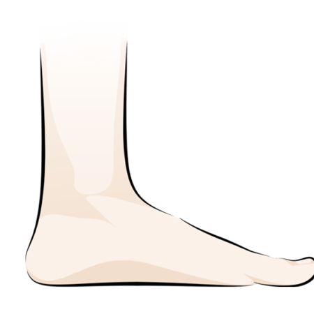
Flat feet are feet where the inner arch is low or absent when you stand. They are extremely common, usually harmless, and only sometimes painful. The confusing part is the vocabulary: flat feet, pes planus, fallen arches and over-pronation are used as if they mean the same thing, but each describes something slightly different.
This guide untangles those terms, then covers who gets flat feet and why, when they matter, how health professionals assess them, and every main treatment, from exercise and footwear to surgery for children and adults. It draws on published research throughout, with links so you can read the studies yourself.
If you are mainly looking for footwear support, see our separate page on insoles for flat feet and fallen arches.
Flat feet, pes planus, fallen arches and over-pronation: what’s the difference?
All four terms describe a foot whose inner (medial longitudinal) arch sits low when it takes weight. They differ in what they emphasize: a shape, a medical label, a change over time, or a movement.
| Term | What it describes | Shape or movement? | Typical use |
|---|---|---|---|
| Flat feet | A low or absent arch when standing | Shape | Everyday language; covers every type |
| Pes planus (also “pes planovalgus”) | The medical name for flat feet; “valgus” adds that the heel tilts outward | Shape | Doctors, podiatrists and research papers |
| Fallen arches | An arch that used to be higher and has dropped, usually in adulthood | Shape that has changed | Adults whose feet have flattened with age, weight, pregnancy or a tendon problem |
| Over-pronation | The foot rolling inward too far, or for too long, as it takes weight | Movement | Running shops, sports medicine, gait analysis |
Pronation itself is normal
Pronation is the natural inward roll of the foot that absorbs shock each time it lands. Everyone pronates. A study that pooled foot-posture measurements from 619 healthy adults found that a slightly pronated foot is the normal resting position (Redmond, Crane & Menz, 2008). “Over-pronation” only describes movement that goes beyond the usual range, and experts disagree on exactly where that line sits.
What they have in common
Whatever the label, the same structures are involved: the heel bone tips inward, the arch lowers, the forefoot turns slightly outward, and the soft tissues that hold the arch up (the tibialis posterior tendon, the plantar fascia, the spring ligament and the small muscles of the sole) carry more load. That is why the same treatments, from strengthening to footwear, come up for all of them.
Flexible vs rigid flat feet
The most useful distinction in practice is not the name but whether the foot is flexible or rigid.
- Flexible flat feet: the arch reappears when you sit, stand on your toes or lift your big toe. This is by far the most common type and is often painless.
- Rigid flat feet: the arch stays flat in every position and the foot is stiff. This is much less common and usually has a structural cause, such as two bones that never separated (tarsal coalition), arthritis or an old injury. It more often needs medical assessment.
Adult-acquired flat foot (progressive collapsing foot deformity)
When an adult foot gradually flattens, often on one side, the cause is frequently a failing tibialis posterior tendon. In 2020, an international expert panel renamed this condition progressive collapsing foot deformity (PCFD), because it involves the whole foot and ankle, not just the arch, and because it tends to worsen without treatment (Myerson et al., 2020). You may still see the older names: adult-acquired flatfoot deformity (AAFD) and posterior tibial tendon dysfunction (PTTD).
How common are flat feet?
Very common, though the numbers depend heavily on how “flat” is measured. A 2023 systematic review that pooled 12 population studies covering 16,000 people put the overall figure at about 15.6%. Flat feet were more common in boys and men, in children aged 3 to 5 and 11 to 17, in people of Asian background and in people living with obesity (2023 systematic review).
In older adults the figure is higher. In a random sample of 1,002 adults aged 40 and over in Spain, about 19% had flat footprints, and the chance rose with age and body weight (Pita-Fernández et al., 2015). In that group women were more affected than men, the reverse of what is seen in children, which suggests the causes change across a lifetime.
Flat feet in babies and young children are normal
Babies are born with a fat pad filling the arch and loose ligaments, so almost all toddlers look flat-footed. The arch develops gradually through early childhood. In a study of 835 Austrian children aged 3 to 6, 44% had flexible flat feet: 54% of three-year-olds but only 24% of six-year-olds. Fewer than 1% had a genuinely abnormal flat foot, and the authors judged that more than 90% of the treatments the children were receiving were unnecessary (Pfeiffer et al., 2006).
The practical message for parents: a flexible, painless flat foot in a young child is usually a stage, not a problem. The signs that do deserve a check are listed in the FAQs below.
What causes flat feet?
Flat feet are either something you grow up with or something that develops later. The causes overlap, and many people have more than one.
Lifelong (developmental) flat feet
- Family traits. Arch height runs in families. Many people with lifelong flat feet have a parent with the same foot shape.
- Ligament laxity. People with naturally flexible joints (hypermobility) often have flexible flat feet, because the ligaments that hold the arch give more under load.
- Body weight in childhood. Carrying extra weight while the arch is developing is linked to flatter feet, as the preschool studies above found.
- Tarsal coalition. In about 1% of people, two bones at the back of the foot are joined by bone, cartilage or fibrous tissue from birth (Lemley et al., 2006). It typically causes a stiff, rigid flat foot and pain, often appearing between ages 8 and 16 as the connection hardens.
Flat feet that develop in adulthood (fallen arches)
- Tibialis posterior tendon failure. This tendon runs behind the inner ankle bone and is the main muscle support for the arch. When it degenerates or tears, the arch slowly collapses. It is most common in women over 40, particularly those with obesity, diabetes or high blood pressure.
- Pregnancy. In a University of Iowa study of 49 women, arch height dropped between early pregnancy and five months after birth, and feet became 2 to 10 mm longer. The change appeared to be permanent and was mostly caused by the first pregnancy (Segal et al., 2013). Hormones that loosen ligaments, plus extra weight, are the likely reasons.
- Weight gain and age. Both put more load through the arch over time. A US analysis of national health survey data linked self-reported flat feet with older age, higher body mass index, bunions, hammer toes and arthritis (Shibuya et al., 2010).
- Arthritis. Rheumatoid arthritis in particular can damage the joints and ligaments of the midfoot.
- Injury. Fractures or ligament injuries in the midfoot (for example a Lisfranc injury) can leave the arch lower.
- Nerve damage. Diabetic neuropathy can, in severe cases, lead to Charcot foot, where the bones of the arch weaken and collapse. This is a medical emergency.
Symptoms: do flat feet actually cause problems?
Many people with flat feet never have symptoms. When they do, the most common complaints are:
- aching or tiredness in the arch, heel or calf after standing or walking
- pain along the inner ankle, sometimes with swelling (a sign to check the tibialis posterior tendon)
- heel pain or plantar fasciitis
- shin pain during or after running
- shoes wearing down quickly on the inner edge
- in rigid flat feet, stiffness and pain on the outer side of the foot, sometimes with repeated ankle sprains
What the research says about flat feet and injury
The link between foot posture and injuries further up the leg is real but smaller than often claimed. A systematic review of 21 prospective studies involving 6,228 people found strong evidence that a pronated foot posture is a risk factor for medial tibial stress syndrome (“shin splints”), and very limited evidence for kneecap pain. The effects were small, and no link was found with foot and ankle injuries, bone stress injuries or general overuse injuries (Neal et al., 2014).
In other words, a flat foot raises the odds of certain problems a little, but it is one factor among many, alongside training load, body weight, footwear and strength. It is not a reason to treat a painless flat foot.
Warning signs to get checked
See a health professional if one foot is becoming flatter than the other, you have pain and swelling behind the inner ankle bone, you can no longer rise onto the toes of one foot, the foot is stiff and painful, or you have diabetes and notice any change in foot shape, warmth or redness.
How are flat feet diagnosed?
Most flat feet are diagnosed by looking at and handling the foot. Scans are only needed when the foot is rigid, painful in an unusual way, or changing.
Simple tests in the clinic
- Standing inspection. Viewed from behind, a flat foot often shows the heel tilting outward and more of the outer toes visible beside the ankle (the “too many toes” sign). A difference between the two feet is especially important.
- Tiptoe test. On standing on tiptoe, a flexible flat foot regains its arch and the heel swings inward. If neither happens, the foot is rigid.
- Single-leg heel raise. Being unable to rise onto the toes of one foot, or the heel not turning inward as you rise, points to a weak tibialis posterior tendon.
- Big-toe lift (Jack’s test). Lifting the big toe while you stand tightens the plantar fascia and raises the arch in a flexible foot.
- Navicular drop. The clinician marks a bone on the inner arch and measures how far it drops between sitting and standing. A larger drop means more arch collapse under load.
- Foot Posture Index (FPI). A six-item checklist scored from −12 (strongly supinated) to +12 (strongly pronated). Normative data from 1,648 observations show that the average healthy adult scores about +4, and that children and older adults naturally score higher (Redmond, Crane & Menz, 2008).
- Footprint measures. A footprint taken on a pressure mat or podoscope can be analyzed with simple angles. In the Spanish study of adults over 40, one of these, Clarke’s angle, was highly accurate at identifying clinically diagnosed flat feet (Pita-Fernández et al., 2015).
Many clinicians also watch you walk or run, sometimes on video, to see how the foot moves rather than just how it looks standing still.
Imaging
- Weight-bearing X-rays measure the angles between the bones of the arch and show arthritis. They must be taken standing; a flat foot can look normal lying down.
- CT scans are the best way to confirm a tarsal coalition. Weight-bearing CT is increasingly used to plan surgery for adult-acquired flat foot.
- Ultrasound or MRI shows the condition of the tibialis posterior tendon and the spring ligament.
How are flat feet treated?
Treatment depends on whether the foot hurts, whether it is flexible, and whether it is changing. A flat foot that is flexible and painless needs no treatment at all, in children or adults.
1. Exercise and strengthening
The small muscles in the sole of the foot help hold the arch up, and they can be trained like any other muscle. The best-studied exercise is the “short-foot” exercise: with the foot flat on the floor, you draw the ball of the foot toward the heel to lift the arch, without curling the toes, and hold for a few seconds.
- In a four-week training program, short-foot exercise reduced how far the arch dropped on standing and increased arch height (Mulligan & Cook, 2013).
- In a pilot randomized trial in people with flat feet, eight weeks of intrinsic foot-muscle training three times a week improved standing foot alignment and arch behaviour during walking (Okamura et al., 2020).
- In a small trial of 14 university students with flexible flat feet, short-foot exercise improved arch drop and balance more than arch-support insoles alone (Kim & Kim, 2016).
The studies are small, but the exercises are safe, free and take a few minutes a day. A typical program also includes:
- Calf stretches, because a tight calf forces the foot to flatten to let the shin move forward over it.
- Heel raises, progressing to single-leg raises, to strengthen the calf and the tibialis posterior tendon.
- Balance work, such as standing on one foot while keeping the arch lifted.
A physiotherapist can check your technique and progress the program.
2. Footwear, weight and activity
Shoes with a firm heel counter, a stable midsole and room for the toes give a flat foot a solid base. If you are carrying extra weight, losing some reduces the load on the arch every step. When symptoms flare, swapping some running or court time for cycling or swimming lets irritated tissues settle.
3. Insoles and orthotics
Arch-support insoles reduce the strain on the tissues that hold up the arch and are usually the next step after exercise and footwear. We cover how to choose them in our guide to insoles for flat feet.
4. Treatment for adult-acquired flat foot
Early tibialis posterior tendon problems are usually treated without surgery: a period of protection in a walking boot or brace if the tendon is very sore, followed by a structured strengthening program, supportive footwear and orthotics. An ankle brace (ankle-foot orthosis) can help people who are not candidates for surgery.
5. Surgery
Surgery is only considered when a flat foot is painful and has not improved with months of non-surgical care, or when the deformity is rigid or progressing. The operation depends on age and the cause.
| Who | Procedure | What it involves | What the research shows |
|---|---|---|---|
| Children with painful flexible flat feet | Subtalar arthroereisis | A small implant or screw is placed in the side of the hindfoot to block excessive inward roll | A review of 24 studies (2,550 feet) reported favourable outcomes and high satisfaction (Smith et al., 2021); a long-term review of 1,536 feet found implant problems in about 3% and wound problems in about 1% (Smolle et al., 2022). All the studies were case series |
| Children and adults with flexible flat feet | Lateral column lengthening | The heel bone is cut and lengthened with a bone graft to restore the arch | Corrects the arch shape more than arthroereisis on X-ray, but is a bigger operation with longer recovery |
| Children with tarsal coalition | Coalition resection | The bony or fibrous bridge is removed and tissue placed in the gap | Usually offered when pain persists despite a period in a cast or boot |
| Adults with flexible adult-acquired flat foot | Calcaneal osteotomy + tendon transfer | The heel bone is shifted inward and a neighbouring toe tendon (flexor digitorum longus) replaces the failed tibialis posterior | In 31 patients followed for about 15 years, foot scores rose from 48 to 90 out of 100 and 87% were pain-free and functioning well (Chadwick et al., 2015) |
| Adults with rigid or arthritic flat feet | Fusion (arthrodesis) | Painful joints in the hindfoot are fused in a corrected position | Reliable pain relief, at the cost of permanent stiffness in the fused joints |
Recovery from adult flat-foot reconstruction is long. Expect several weeks without putting weight on the foot, then a walking boot, and six to twelve months before the final result. Your surgeon will give you a plan specific to the operation.
Getting help for flat feet in Canada
- Family doctor: first assessment, X-ray requisitions and referral to an orthopaedic surgeon if needed.
- Physiotherapist: strengthening, stretching and balance programs; often the best first visit for painful flexible flat feet.
- Podiatrist or chiropodist (depending on your province): foot assessment, gait analysis and orthotics. In some provinces podiatrists also perform foot surgery.
- Canadian Certified Pedorthist (pedorthic.ca): footwear and orthotic assessment and fitting.
- Orthopaedic foot and ankle surgeon: by referral, for rigid, progressive or surgically treatable flat feet. Surgery in hospital is covered by your provincial health plan; physiotherapy, podiatry, chiropody and pedorthic visits usually are not, but many extended health plans include them.
For children, start with your family doctor or paediatrician, particularly if the foot is stiff, painful, different from the other side, or your child tires much sooner than friends.
FAQs about flat feet
1. Are flat feet hereditary?
Often, yes. Arch height and joint flexibility both run in families, so many people with lifelong flat feet have a parent with similar feet. Tarsal coalition also has a family link. Flat feet that develop in adulthood, by contrast, are usually caused by tendon problems, weight, pregnancy or arthritis rather than genes.
2. When should I worry about my child’s flat feet?
Most young children have flat feet, and the arch usually develops by around age six to ten. Have your child checked if the foot is stiff, painful or very different from the other side; if the arch doesn’t reappear when they stand on tiptoe; if they tire or limp much sooner than other children; or if the flat foot appears or worsens after age ten.
3. Can exercises give me an arch?
Exercises can raise the arch slightly and, more importantly, improve how well it holds up under load. Studies of short-foot exercise show less arch drop and better balance within four to eight weeks. They won’t turn a naturally flat foot into a high-arched one, but symptoms often improve because the foot is stronger.
4. Do flat feet get worse with age?
They can. Ligaments and tendons lose some stiffness with age, weight often increases, and the tibialis posterior tendon can wear. A foot that flattens noticeably over months, especially on one side, should be assessed, because adult-acquired flat foot tends to progress without treatment.
5. Is flattening of the feet during pregnancy permanent?
For many women, partly yes. A study following 49 women found that arch height dropped and feet lengthened by 2 to 10 mm between early pregnancy and five months after birth, with no recovery, and most of the change came with the first pregnancy. Supportive footwear during pregnancy and foot-strengthening exercises afterward are sensible, though neither has been proven to prevent the change.
6. Is walking barefoot good or bad for flat feet?
It depends on your symptoms. For a painless flexible flat foot, time barefoot on varied surfaces such as grass or sand works the small foot muscles and is generally fine. If your feet already ache, long periods barefoot on hard floors usually make things worse; build up gradually and wear supportive shoes for long days.
7. Are flat feet and plantar fasciitis related?
They often occur together. When the arch lowers, the plantar fascia, the thick band along the sole, is stretched more with every step, which can irritate its attachment at the heel. People with high arches get plantar fasciitis too, though, so flat feet are one risk factor rather than the cause. Read more in our plantar fasciitis insoles guide.
References
- Redmond AC, Crane YZ, Menz HB. Normative values for the Foot Posture Index. J Foot Ankle Res. 2008;1:6. doi:10.1186/1757-1146-1-6
- Myerson MS, Thordarson DB, Johnson JE, et al. Classification and nomenclature: progressive collapsing foot deformity. Foot Ankle Int. 2020;41(10):1271–1276. doi:10.1177/1071100720950722
- Prevalence and clinical factors associated with pes planus among children and adults: a population-based synthesis and systematic review. J Foot Ankle Surg. 2023;62(5):899–903. doi:10.1053/j.jfas.2023.05.007
- Pita-Fernández S, et al. Validity of footprint analysis to determine flatfoot using clinical diagnosis as the gold standard in a random sample aged 40 years and older. J Epidemiol. 2015;25(2):148–154. doi:10.2188/jea.JE20140082
- Pfeiffer M, Kotz R, Ledl T, Hauser G, Sluga M. Prevalence of flat foot in preschool-aged children. Pediatrics. 2006;118(2):634–639. doi:10.1542/peds.2005-2126
- Lemley F, et al. Current concepts review: tarsal coalition. Foot Ankle Int. 2006;27(12):1163–1169. PubMed 17207452
- Segal NA, Boyer ER, Teran-Yengle P, et al. Pregnancy leads to lasting changes in foot structure. Am J Phys Med Rehabil. 2013;92(3):232–240. doi:10.1097/PHM.0b013e31827443a9
- Shibuya N, Jupiter DC, Ciliberti LJ, VanBuren V, La Fontaine J. Characteristics of adult flatfoot in the United States. J Foot Ankle Surg. 2010;49(4):363–368. Record
- Neal BS, Griffiths IB, Dowling GJ, et al. Foot posture as a risk factor for lower limb overuse injury: a systematic review and meta-analysis. J Foot Ankle Res. 2014;7:55. doi:10.1186/s13047-014-0055-4
- Mulligan EP, Cook PG. Effect of plantar intrinsic muscle training on medial longitudinal arch morphology and dynamic function. Manual Therapy. 2013;18(5):425–430. doi:10.1016/j.math.2013.02.007
- Okamura K, Fukuda K, Oki S, et al. Effects of plantar intrinsic foot muscle strengthening exercise on static and dynamic foot kinematics: a pilot randomized controlled single-blind trial in individuals with pes planus. Gait Posture. 2020;75:40–45. doi:10.1016/j.gaitpost.2019.09.030
- Kim EK, Kim JS. The effects of short foot exercises and arch support insoles on improvement in the medial longitudinal arch and dynamic balance of flexible flatfoot patients. J Phys Ther Sci. 2016;28(11):3136–3139. doi:10.1589/jpts.28.3136
- Smith C, et al. Subtalar arthroereisis for the treatment of the symptomatic paediatric flexible pes planus: a systematic review. EFORT Open Rev. 2021;6(2):118–129. doi:10.1302/2058-5241.6.200076
- Smolle MA, Svehlik M, Regvar K, Leithner A, Kraus T. Long-term clinical and radiological outcomes following surgical treatment for symptomatic pediatric flexible flat feet: a systematic review. Acta Orthop. 2022;93:367–374. Full text
- Chadwick C, Whitehouse SL, Saxby TS. Long-term follow-up of flexor digitorum longus transfer and calcaneal osteotomy for stage II posterior tibial tendon dysfunction. Bone Joint J. 2015;97-B(3):346–352. doi:10.1302/0301-620X.97B3.34386
- Pedorthic Association of Canada. Find a Canadian Certified Pedorthist
